Types of Scans for Diagnosis
Types of Scans for Diagnosis
Had your doctor asked you to get an X-Ray or MRI or any other scan? Do you know why these tests are suggested? How do doctors use them to diagnose the illness? Medical Scans are used to diagnose any ailment from torn ligaments to the tumor. There are various types of scans and each of them works differently. Some use radiations, some magnets, some radio waves, and some sound waves.
So, let’s read about types of scans:
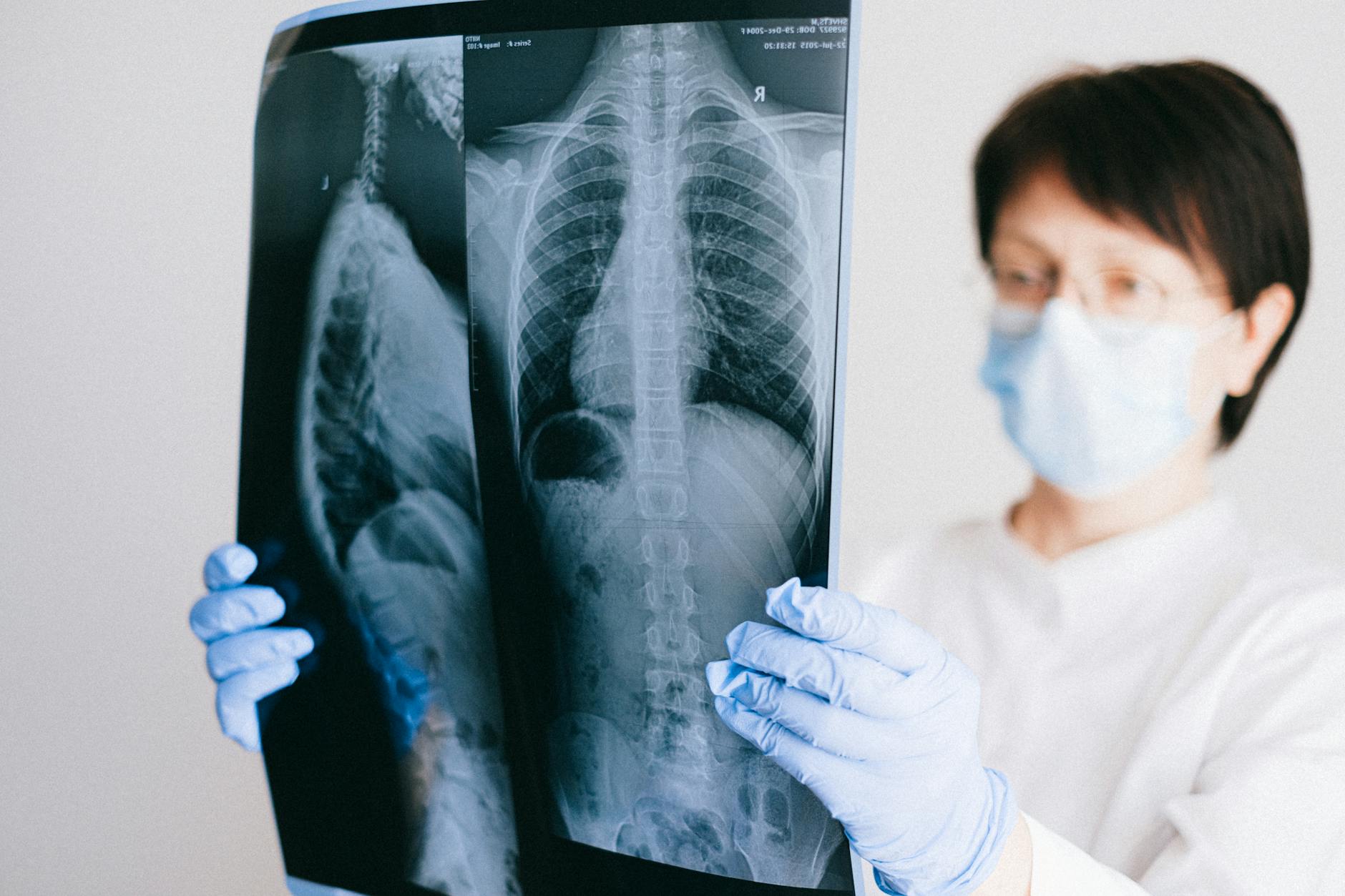
X-Rays –
These are the most commonly used scans that create images of the inner body parts. X-Rays are majorly used for detecting fractures (broken bones), detecting pneumonia, tumors, cancers, etc. They travel easily through air-filled cavities like lungs and less radiologically dense tissues like fat and muscle.
The parts of the body are displayed in white and black shades because the different amount of radiation is absorbed by different tissues. Bones look white because calcium absorbs x-rays the most, the air absorbs the least x-ray so lungs look black and fat and other soft tissues look grey because they absorb fewer x-rays.
CT Scan –
CT Scan is a Computerized Tomography that combines the images of the X-Ray taken from various angles to create a two-dimensional image of bones, blood vessels, and soft tissues.
It is also known as Computerized Axial Tomography (CAT) and is used in emergencies when the doctor wants to know if there is any life-threatening condition.
It has many uses but it is well suited for examining people having internal injuries during car accidents or any kind of trauma. It is painless and with the development of new machines, it takes less time. Typically, the whole process is around 30 minutes.
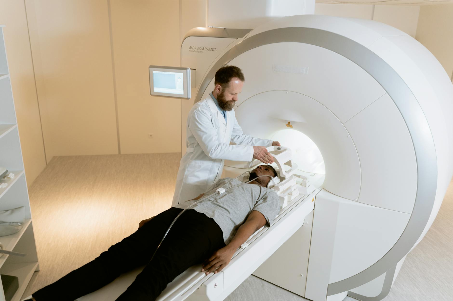
MRI –
MRI is a Magnetic Resonance Imaging scan that helps to detect any brain injuries, nerve injuries, tumors, and even the reason behind a headache. It makes use of the strong magnet and radio waves and no radiation is involved in it.
These affect the atoms that are present in the water molecules of your body’s tissue. When the radio waves are stopped, the energy is released by the atoms which in turn is detected by the MRI machine.
There is hardly any side effect of MRI other than headache or nausea. The time taken to perform this scan may vary from 10 minutes to 1 hour. You need to lie motionless and the technicians may offer you an earplug as it can be loud.
For example, you can visit TopMri as it helps to save money on imaging services by booking faster appointments at diagnostic centers across the UK.
Ultrasound –
Ultrasound is widely used to examine various parts of the body like the liver, kidneys, gallbladder, etc. It is majorly used at the time of pregnancy to detect the development of the baby inside the womb.
It is also used to identify any injury in the shoulder, knee, and ankle, and can also be used to check the flow of the blood and any blocked vessels.
This scan is performed with the help of a transducer and a water-based gel. The sonographer moves the transducer over the skin and images are generated using high-frequency sound waves.
The doctor may ask you to keep your bladder full or even too fast before the ultrasound. It takes around half an hour to an hour and does not require any kind of medication.
PET –
PET is Positron Emission Tomography which is mainly used in cardiology, neurology, infection, and inflammation, and even in planning treatments and surgeries. It gives two types of information from a single examination – functional and structural as it uses two different techniques in one scanner.
It shows the changes in the tissues in which glucose is the main source of energy, for example, the heart muscle or the brain.
A radioactive tracer is injected into the body which gives off the gamma rays. The drug travels to the places where glucose is stored in the form of energy and shows cancer which in turn is detected by PET.
Fluoroscopy –
It is a medical imagining technique in which the image of the continuous x-ray is displayed on the screen. It is used to obtain real-time moving images of the patient’s internal body with the use of an x-ray and fluorescent screen.
In this procedure, an x-ray beam is passed through the body. It is recorded to play at a later stage for more detailed analysis.
Bone Densitometry (DEXA) –
It is a scanning technique that is used to gauge the mineral bone density and can diagnose any kind of condition like arthritis, bowel disorder, hormonal disorder, etc.
It makes use of radionuclides which are radioactive chemicals. These are injected or breathed or swallowed into the body to take the bone’s images.
The calculations that are performed give an almost exact amount of mineral that is present in the bone. Then, the comparison is done with people of similar age, sex, weight, etc.
Conclusion
Scientists are working hard to find other ways of scanning in which results are quick and radiation is less. They are also working to develop an advanced technique to detect some conditions like infections that are hidden deep inside the body. There are various types of scans depending on the kind of illness. In this article, we have discussed a few types of scans that are used to diagnose diseases.
Source
https://www.alliancemedical.co.uk/what-we-do/patient-services/types-of-scan
https://www.dignityhealth.org/articles/types-of-scans-and-medical-imaging
https://newsinhealth.nih.gov/2019/11/medical-scans-explained